3D-printed calcium magnesium silicates: A mini-review

  • Aidin Doroudi 1
  • Preeti Lata Mahapatra 2
  • Fatemeh Bakhshi 3
  • 1 Polymer and Color Engineering Department, Amirkabir University of Technology, Tehran, Iran
  • 2 School of Nano Science and Technology, Indian Institute of Technology, Kharagpur, West Bengal-721302, India
  • 3 Polymer and color Engineering department, Amirkabir University of Technology, Tehran, Iran

Abstract

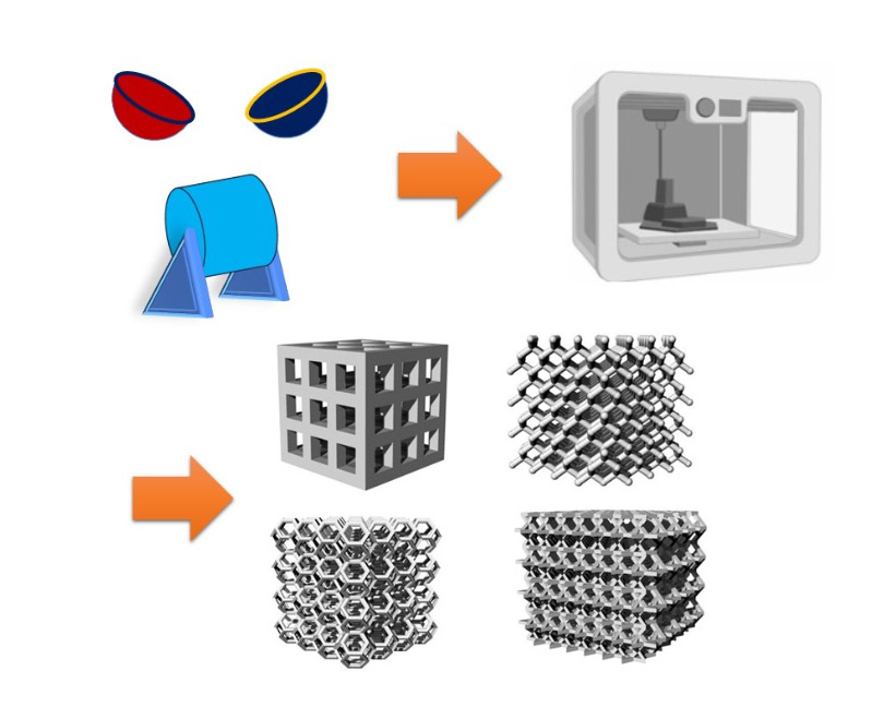
Calcium magnesium silicates (CMS) represent a class of minerals with diverse applications in fields ranging from geology to materials science. With the advent of additive manufacturing technologies, particularly 3D printing, novel opportunities have emerged for the synthesis and utilization of CMS-based materials. In this mini-review, we provide a thorough overview of recent advancements in the 3D printing of CMS compounds, including diopside (DPS), bredigite (BR), and akermanite (AKT). We discuss the synthesis methods, properties, and potential applications of 3D-printed CMS materials, with a focus on their role in biomedical applications. Furthermore, we highlight challenges and prospects in the field, emphasizing the importance of continued research and innovation in harnessing the full potential of 3D-printed CMS materials.

Downloads

Download data is not yet available.
Keywords: Calcium magnesium silicates, 3D printing, Synthesis methods, Bioceramics, Additive manufacturing

References

[1] L.L. Hench, R.J. Splinter, W.C. Allen, T.K. Greenlee, Bonding mechanisms at the interface of ceramic prosthetic materials, J. Biomed. Mater. Res. 5 (1971) 117–141. https://doi.org/10.1002/jbm.820050611.
[2] C. Wu, J. Chang, Degradation, bioactivity, and cytocompatibility of diopside, akermanite, and bredigite ceramics, J. Biomed. Mater. Res. B. 83B (2007) 153–160. https://doi.org/10.1002/jbm.b.30779.
[3] S.K. Venkatraman, S. Swamiappan, Review on calcium‐and magnesium‐based silicates for bone tissue engineering applications, J. Biomed. Mater. Res. A. 108 (2020) 1546–1562. https://doi.org/10.1002/jbm.a.36925.
[4] M. Vallet-Regí, Ceramics for medical applications, J. Chem. Soc. Dalton Trans. (2001) 97–108. https://doi.org/10.1039/B007852M.
[5] S. Amirfarhangi, A. Vakili, L. Radfar, N. Tajbakhsh, Golden proportion and facial esthetic, the harmony and surgical considerations: A review, World J. Biol. Pharm. Health Sci. 11 (2022) 018–021. https://doi.org/10.30574/wjbphs.2022.11.1.0097.
[6] A. Shahbaz, N. Tajbakhsh, A. Doroudi, F. Bakhshi, S. Ranjbar, Bredigite-containing materials for regenerative medicine applications: A rapid review, J. Compos. Compd. 5 (2023) 190–199. https://doi.org/10.61186/jcc.5.3.4.
[7] F. Tavangarian, R. Emadi, Improving degradation rate and apatite formation ability of nanostructure forsterite, Ceram. Int. 37 (2011) 2275–2280. https://doi.org/10.1016/j.ceramint.2011.03.022.
[8] S. Ni, J. Chang, In vitro Degradation, Bioactivity, and Cytocompatibility of Calcium Silicate, Dimagnesium Silicate, and Tricalcium Phosphate Bioceramics, J. Biomater. Appl. 24 (2009) 139–158. https://doi.org/10.1177/0885328208094745.
[9] A.H. Shahbaz, M. Esmaeilian, R. NasrAzadani, K. Gavanji, Effect of MgF2 addition on the mechanical properties of hydroxyapatite synthesized via powder metallurgy, J. Compos. Compd. 1 (2019) 16–21. https://doi.org/10.29252/jcc.1.1.3.
[10] H. Ghomi, M. Jaberzadeh, M.H. Fathi, Novel fabrication of forsterite scaffold with improved mechanical properties, J. Alloys Compd. 509 (2011) L63–L68. https://doi.org/10.1016/j.jallcom.2010.10.106.
[11] R. Choudhary, L. Ravi, S. Swamiappan, Silicate ceramics and its composites for hard tissue applications, Eco-Friendly Nano-Hybrid Materials for Advanced Engineering Applications, Apple Academic Press. (2017) 131–182. https://doi.org/10.1201/9781315366531-8.
[12] M. Diba, M. Kharaziha, M.H. Fathi, M. Gholipourmalekabadi, A. Samadikuchaksaraei, Preparation and characterization of polycaprolactone/forsterite nanocomposite porous scaffolds designed for bone tissue regeneration, Compos. Sci. Technol. 72 (2012) 716–723. https://doi.org/10.1016/j.compscitech.2012.01.023.
[13] Z. Han, C. Gao, P. Feng, Y. Shen, C. Shuai, S. Peng, Silicon carbide whiskers reinforced akermanite scaffolds for tissue engineering, RSC Adv. 4 (2014) 36868–36874. https://doi.org/10.1039/C4RA07474B.
[14] L. Tingting, D. Youwen, G. Chengde, F. Pei, C. Shuai, P. Shuping, Analysis of 3D printed diopside scaffolds properties for tissue engineering, Mater. Sci. 21 (2015) 590–594. https://doi.org/10.5755/j01.ms.21.4.9845.
[15] A. Dasan, H. Elsayed, J. Kraxner, D. Galusek, E. Bernardo, Hierarchically porous 3D-printed akermanite scaffolds from silicones and engineered fillers, J. Eur. Ceram. Soc. 39 (2019) 4445–4449. https://doi.org/10.1016/j.jeurceramsoc.2019.06.021.
[16] Z. Han, P. Feng, C. Gao, Y. Shen, C. Shuai, S. Peng, Microstructure, mechanical properties and in vitro bioactivity of akermanite scaffolds fabricated by laser sintering, Bio-Med. Mater. Eng. 24 (2014) 2073–2080. https://doi.org/10.3233/BME-141017.
[17] M.R. Shahrouzifar, E. Salahinejad, Strontium doping into diopside tissue engineering scaffolds, Ceram. Int. 45 (2019) 10176–10181. https://doi.org/10.1016/j.ceramint.2019.02.067.
[18] S. Pang, D. Wu, F. Kamutzki, J. Kurreck, A. Gurlo, D.A.H. Hanaor, High performing additively manufactured bone scaffolds based on copper substituted diopside, Mater. Des. 215 (2022) 110480. https://doi.org/10.1016/j.matdes.2022.110480.
[19] Z. Ba, Z. Chen, Y. Huang, D. Feng, Q. Zhao, et al., Nanoporous diopside modulates biocompatibility, degradability and osteogenesis of bioactive scaffolds of gliadin-based composites for new bone formation, Int. J. Nanomed. (2018) 3883–3896. https://doi.org/10.2147/IJN.S162262.
[20] A. Dasan, J. Kraxner, L. Grigolato, G. Savio, H. Elsayed, et al., 3D Printing of Hierarchically Porous Lattice Structures Based on Åkermanite Glass Microspheres and Reactive Silicone Binder, J. Funct. Biomater. 13 (2022) 8. https://doi.org/10.3390/jfb13010008.
[21] M. Hafezi-Ardakani, F. Moztarzadeh, M. Rabiee, A.R. Talebi, M. Abasi-shahni, et al., Sol-gel synthesis and apatite-formation ability of nanostructure merwinite (Ca3MgSi2O8) as a novel bioceramic, J. Ceram. Process. Res. 11 (2010) 765–768
[22] M.S. Collin, S.K. Venkatraman, M. Sriramulu, S. Shanmugam, E.A. Drweesh, et al., Solution combustion synthesis of functional diopside, akermanite, and merwinite bioceramics: Excellent biomineralization, mechanical strength, and antibacterial ability, Mater. Today Commun. 27 (2021) 102365. https://doi.org/10.1016/j.mtcomm.2021.102365.
[23] M. Ansari, F. Malmir, A. Salati, Preparation and Characterization of Akermanite/Merwinite Scaffolds for Bone Tissue Repair, J. Biomim. Biomater. Biomed. Eng. 44 (2020) 73–81. https://doi.org/10.4028/www.scientific.net/JBBBE.44.73.
[24] M. Razavi, M. Fathi, O. Savabi, D. Vashaee, L. Tayebi, In vivo biocompatibility of Mg implants surface modified by nanostructured merwinite/PEO, J. Mater. Sci. Mater. Med. 26 (2015) 184. https://doi.org/10.1007/s10856-015-5514-3.
[25] Y. Hosseini, R. Emadi, M. Kharaziha, Surface modification of PCL-diopside fibrous membrane via gelatin immobilization for bone tissue engineering, Mater. Chem. Phys. 194 (2017) 356–366. https://doi.org/10.1016/j.matchemphys.2017.03.051.
[26] J. Ou, G.F. Yin, D.L. Zhou, X.C. Chen, Y.D. Yao, et al., Preparation of Merwinite with Apatite-Forming Ability by Sol-Gel Process, Key Eng. Mater. 330–332 (2007) 67–70. https://doi.org/10.4028/www.scientific.net/KEM.330-332.67.
[27] C. Shuai, Z. Han, P. Feng, C. Gao, T. Xiao, S. Peng, Akermanite scaffolds reinforced with boron nitride nanosheets in bone tissue engineering, J. Mater. Sci: Mater. Med. 26 (2015) 188. https://doi.org/10.1007/s10856-015-5513-4.
[28] G. Birhanu, M. Doosti-Telgerd, A. Zandi-Karimi, Z. Karimi, M. Porgham Daryasari, et al., Enhanced proliferation and osteogenic differentiation of mesenchymal stem cells by diopside coated Poly-L-lactic Acid-Based nanofibrous scaffolds, Int. J. Polym. Mater. Polym. Biomater. 71 (2022) 707–716. https://doi.org/10.1080/00914037.2021.1879078.
[29] S.K. Venkatraman, R. Choudhary, G. Krishnamurithy, H.R. Balaji Raghavendran, M.R. Murali, et al., Comparative investigation on antibacterial, biological and mechanical behaviour of monticellite and diopside derived from biowaste for bone regeneration, Mater. Chem. Phys. 286 (2022) 126157. https://doi.org/10.1016/j.matchemphys.2022.126157.
[30] H. Mohammadi, Y.M.B. Ismail, K.A. Shariff, A.F.M. Noor, Effect of substitutional Strontium on Mechanical Properties of Akermanite Ceramic Prepared by Solid-State Sintering, Mater. Today: Proc. 17 (2019) 929–936. https://doi.org/10.1016/j.matpr.2019.06.393.
[31] A.K. Sharafabadi, M. Abdellahi, A. Kazemi, A. Khandan, N. Ozada, A novel and economical route for synthesizing akermanite (Ca2MgSi2O7) nano-bioceramic, Mater. Sci. Eng: C. 71 (2017) 1072–1078. https://doi.org/10.1016/j.msec.2016.11.021.
[32] R. Choudhary, S. Koppala, S. Swamiappan, Bioactivity studies of calcium magnesium silicate prepared from eggshell waste by sol–gel combustion synthesis, J. Asian Ceram. Soc. 3 (2015) 173–177. https://doi.org/10.1016/j.jascer.2015.01.002.
[33] S. Yamamoto, N. Kawamura, T. Nonami, Diopside Synthesized by Sol-gel Method as Phosphorus Adsorption Material: Evaluation of Apatite Deposition in Pseudo Body Solution, Trans. Mater. Res. Soc. Jpn. 44 (2019) 17–23. https://doi.org/10.14723/tmrsj.44.17.
[34] R. Negrea, C. Busuioc, I. Constantinoiu, D. Miu, C. Enache, et al., Akermanite-based coatings grown by pulsed laser deposition for metallic implants employed in orthopaedics, Surf. Coat. Technol. 357 (2019) 1015–1026. https://doi.org/10.1016/j.surfcoat.2018.11.008.
[35] A.-E. Alecu, C.-C. Costea, V.-A. Surdu, G. Voicu, S.-I. Jinga, C. Busuioc, Processing of Calcium Magnesium Silicates by the Sol–Gel Route, Gels. 8 (2022) 574. https://doi.org/10.3390/gels8090574.
[36] X. Wang, L. Zhang, X. Ke, J. Wang, G. Yang, et al., 45S5 Bioglass analogue reinforced akermanite ceramic favorable for additive manufacturing mechanically strong scaffolds, RSC Adv. 5 (2015) 102727–102735. https://doi.org/10.1039/c5ra19272b.
[37] P.L. Mahapatra, R. Tromer, A. Jayakumar, G. Costin, B. Lahiri, et al., 3D-printed flexible energy harvesting devices designed using non-layered two-dimensional natural tourmaline silicates, J. Mater. Chem. C. 12 (2024) 3418–3429. https://doi.org/10.1039/D3TC04167K.
[38] Q. Fu, E. Saiz, M.N. Rahaman, A.P. Tomsia, Bioactive glass scaffolds for bone tissue engineering: state of the art and future perspectives, Mater. Sci. Eng: C. 31 (2011) 1245–1256. https://doi.org/10.1016/j.msec.2011.04.022.
[39] M. Abdellahi, A. Najafinezhad, H. Ghayour, S. Saber-Samandari, A. Khandan, Preparing diopside nanoparticle scaffolds via space holder method: Simulation of the compressive strength and porosity, J. Mech. Behav. Biomed. Mater. 72 (2017) 171–181. https://doi.org/10.1016/j.jmbbm.2017.05.004.
[40] H. Ghomi, R. Emadi, S.H. Javanmard, Fabrication and characterization of nanostructure diopside scaffolds using the space holder method: Effect of different space holders and compaction pressures, Mater. Des. 91 (2016) 193–200. https://doi.org/10.1016/j.matdes.2015.11.078.
[41] H. Ghomi, R. Emadi, Fabrication of bioactive porous bredigite (Ca7MgSi4O16) scaffold via space holder method, International J. Mater. Res. 109 (2018) 257–264. https://doi.org/10.3139/146.111597.
[42] A. Mansourighasri, N. Muhamad, A.B. Sulong, Processing titanium foams using tapioca starch as a space holder, J. Mater. Proc. Technol. 212 (2012) 83–89. https://doi.org/10.1016/j.jmatprotec.2011.08.008.
[43] M. Kouhi, M. Fathi, V. Jayarama Reddy, S. Ramakrishna, Bredigite Reinforced Electrospun Nanofibers for Bone Tissue Engineering, Mater. Today: Proc. 7 (2019) 449–454. https://doi.org/10.1016/j.matpr.2018.11.108.
[44] M. Kouhi, M. Fathi, M.P. Prabhakaran, M. Shamanian, S. Ramakrishna, Poly L lysine-modified PHBV based nanofibrous scaffolds for bone cell mineralization and osteogenic differentiation, Appl. Surf. Sci. 457 (2018) 616–625. https://doi.org/10.1016/j.apsusc.2018.06.239.
[45] Y. Hosseini, R. Emadi, M. Kharaziha, A. Doostmohammadi, Reinforcement of electrospun poly(ε-caprolactone) scaffold using diopside nanopowder to promote biological and physical properties, J. Appl. Polym. Sci. 134 (2017). https://doi.org/10.1002/app.44433.
[46] A. Khademhosseini, R. Langer, A decade of progress in tissue engineering, Nat. Protoc. 11 (2016) 1775–1781. https://doi.org/10.1038/nprot.2016.123.
[47] B. Baumann, T. Jungst, S. Stichler, S. Feineis, O. Wiltschka, et al., Control of Nanoparticle Release Kinetics from 3D Printed Hydrogel Scaffolds, Angew. Chem. Int. Ed. 56 (2017) 4623–4628. https://doi.org/10.1002/anie.201700153.
[48] N. Tajbakhsh, F. Delpisheh, N. Ghadimi, S. Ansari, Smile management: White esthetic, pink esthetic and facial attractiveness, a review of literature, Open Access Res. J. Biol. Pharm. 05 (2022) 046–050. https://doi.org/10.53022/oarjbp.2022.5.2.0054.
[49] Y. Zhang, D. Zhai, M. Xu, Q. Yao, H. Zhu, et al., 3D-printed bioceramic scaffolds with antibacterial and osteogenic activity, Biofabrication. 9 (2017) 025037. https://doi.org/10.1088/1758-5090/aa6ed6.
[50] I. Sabree, J.E. Gough, B. Derby, Mechanical properties of porous ceramic scaffolds: Influence of internal dimensions, Ceram. Int. 41 (2015) 8425–8432. https://doi.org/10.1016/j.ceramint.2015.03.044.
[51] R. Wang, P. Zhu, W. Yang, S. Gao, B. Li, Q. Li, Direct-writing of 3D periodic TiO2 bio-ceramic scaffolds with a sol-gel ink for in vitro cell growth, Mater. Des. 144 (2018) 304–309. https://doi.org/10.1016/j.matdes.2018.02.040.
[52] X. Zhang, Y. Li, X. Dong, H. Wang, B. Chen, et al., 3D-printed bioactive ceramic scaffolds with MoSe2 nanocrystals as photothermal agents for bone tumor therapy, RSC Adv. 12 (2022) 30588–30597. https://doi.org/10.1039/D2RA02942A.
[53] C. Qin, D. Che, D. Liu, Z. Zhang, Y. Feng, Preparation and characterization of different micro/nano structures on the surface of bredigite scaffolds, Sci. Rep. 13 (2023) 9072. https://doi.org/10.1038/s41598-023-36382-z.
[54] D. Liu, X. Zhou, F. Wang, Y. Feng, Y. Shi, Research and analysis of the properties of bredigite-based 3D-printed bone scaffolds, IJB. 9 (2023) 708. https://doi.org/10.18063/ijb.708.
[55] Y. Xuan, L. Li, C. Zhang, M. Zhang, J. Cao, Z. Zhang, The 3D-Printed Ordered Bredigite Scaffold Promotes Pro-Healing of Critical-Sized Bone Defects by Regulating Macrophage Polarization, Int. J. Nanomed. 18 (2023) 917–932. https://doi.org/10.2147/IJN.S393080.
[56] C. Wu, Y. Ramaswamy, H. Zreiqat, Porous diopside (CaMgSi2O6) scaffold: A promising bioactive material for bone tissue engineering, Acta Biomater. 6 (2010) 2237–2245. https://doi.org/10.1016/j.actbio.2009.12.022.
[57] J. Wei, F. Chen, J.-W. Shin, H. Hong, C. Dai, et al., Preparation and characterization of bioactive mesoporous wollastonite – Polycaprolactone composite scaffold, Biomaterials. 30 (2009) 1080–1088. https://doi.org/10.1016/j.biomaterials.2008.10.046.
[58] R. Emadi, S.I. Roohani Esfahani, F. Tavangarian, A novel, low temperature method for the preparation of ß-TCP/HAP biphasic nanostructured ceramic scaffold from natural cancellous bone, Mater. Lett. 64 (2010) 993–996. https://doi.org/10.1016/j.matlet.2010.01.085.
[59] L.L. Hench, I. Thompson, Twenty-first century challenges for biomaterials, J. Royal Soc. Interface. 7 (2010) S379–S391. https://doi.org/10.1098/rsif.2010.0151.focus.
[60] I.D. Xynos, A.J. Edgar, L.D.K. Buttery, L.L. Hench, J.M. Polak, Gene-expression profiling of human osteoblasts following treatment with the ionic products of Bioglass® 45S5 dissolution, J. Biomed. Mater. Res. 55 (2001) 151–157. https://doi.org/10.1002/1097-4636(200105)55:2<151::AID-JBM1001>3.0.CO;2-D.
[61] I.D. Xynos, A.J. Edgar, L.D.K. Buttery, L.L. Hench, J.M. Polak, Ionic Products of Bioactive Glass Dissolution Increase Proliferation of Human Osteoblasts and Induce Insulin-like Growth Factor II mRNA Expression and Protein Synthesis, Biochem. Biophys. Res. Commun. 276 (2000) 461–465. https://doi.org/10.1006/bbrc.2000.3503.
[62] I.D. Xynos, M.V.J. Hukkanen, J.J. Batten, L.D. Buttery, L.L. Hench, J.M. Polak, Bioglass ®45S5 Stimulates Osteoblast Turnover and Enhances Bone Formation In Vitro: Implications and Applications for Bone Tissue Engineering, Calcif. Tissue Int. 67 (2000) 321–329. https://doi.org/10.1007/s002230001134.
[63] J.R. Jones, Review of bioactive glass: From Hench to hybrids, Acta Biomater. 9 (2013) 4457–4486. https://doi.org/10.1016/j.actbio.2012.08.023.
[64] E. Fiume, J. Barberi, E. Verné, F. Baino, Bioactive Glasses: From Parent 45S5 Composition to Scaffold-Assisted Tissue-Healing Therapies, J. Funct. Biomater. 9 (2018). https://doi.org/10.3390/jfb9010024.
[65] H. Gu, F. Guo, X. Zhou, L. Gong, Y. Zhang, et al., The stimulation of osteogenic differentiation of human adipose-derived stem cells by ionic products from akermanite dissolution via activation of the ERK pathway, Biomaterials. 32 (2011) 7023–7033. https://doi.org/10.1016/j.biomaterials.2011.06.003.
[66] M. Diba, O.-M. Goudouri, F. Tapia, A.R. Boccaccini, Magnesium-containing bioactive polycrystalline silicate-based ceramics and glass-ceramics for biomedical applications, Curr. Opin. Solid State Mater. Sci. 18 (2014) 147–167. https://doi.org/10.1016/j.cossms.2014.02.004.
[67] A. Liu, M. Sun, X. Yang, C. Ma, Y. Liu, et al., Three-dimensional printing akermanite porous scaffolds for load-bearing bone defect repair: An investigation of osteogenic capability and mechanical evolution, J. Biomater. Appl. 31 (2016) 650–660. https://doi.org/10.1177/0885328216664839.
[68] L.D. Shea, D. Wang, R.T. Franceschi, D.J. Mooney, Engineered Bone Development from a Pre-Osteoblast Cell Line on Three-Dimensional Scaffolds, Tissue Eng. 6 (2000) 605–617. https://doi.org/10.1089/10763270050199550.
[69] K.J.L. Burg, S. Porter, J.F. Kellam, Biomaterial developments for bone tissue engineering, Biomaterials. 21 (2000) 2347–2359. https://doi.org/10.1016/S0142-9612(00)00102-2.
[70] B. Bulut, Ş. Duman, Evaluation of mechanical behavior, bioactivity, and cytotoxicity of chitosan/akermanite-TiO2 3D-printed scaffolds for bone tissue applications, Ceram. Int. 48 (2022) 21378–21388. https://doi.org/10.1016/j.ceramint.2022.04.104.
[71] P. Miranda, A. Pajares, E. Saiz, A.P. Tomsia, F. Guiberteau, Mechanical properties of calcium phosphate scaffolds fabricated by robocasting, J. Biomed. Mater. Res. A. 85A (2008) 218–227. https://doi.org/10.1002/jbm.a.31587.
[72] Y. Tanimoto, N. Nishiyama, Preparation and physical properties of tricalcium phosphate laminates for bone-tissue engineering, J. Biomed. Mater. Res. A. 85A (2008) 427–433. https://doi.org/10.1002/jbm.a.31569.
[73] A. Shahbaz, M. Abbasi, H. Sabet, Effect of microstructure on mechanical, electrochemical, and biological properties of Ti/HA surface composites fabricated by FSP method, Mater. Today Commun. 37 (2023) 107305. https://doi.org/10.1016/j.mtcomm.2023.107305.
[74] C. Wu, J. Chang, A review of bioactive silicate ceramics, Biomed. Mater. 8 (2013) 032001. https://doi.org/10.1088/1748-6041/8/3/032001.
[75] W. Zhai, H. Lu, C. Wu, L. Chen, X. Lin, et al., Stimulatory effects of the ionic products from Ca–Mg–Si bioceramics on both osteogenesis and angiogenesis in vitro, Acta Biomater. 9 (2013) 8004–8014. https://doi.org/10.1016/j.actbio.2013.04.024.
[76] X. Dong, A. Heidari, A. Mansouri, W.S. Hao, M. Dehghani, et al., Investigation of the mechanical properties of a bony scaffold for comminuted distal radial fractures: Addition of akermanite nanoparticles and using a freeze-drying technique, J. Mech. Behav. Biomed. Mater. 121 (2021) 104643. https://doi.org/10.1016/j.jmbbm.2021.104643.
[77] X. Ke, L. Zhang, X. Yang, J. Wang, C. Zhuang, et al., Low-melt bioactive glass-reinforced 3D printing akermanite porous cages with highly improved mechanical properties for lumbar spinal fusion, J. Tissue Eng. Regen. Med. 12 (2018) 1149–1162. https://doi.org/10.1002/term.2624.
[78] N.E. Putra, M.A. Leeflang, M. Klimopoulou, J. Dong, P. Taheri, et al., Extrusion-based 3D printing of biodegradable, osteogenic, paramagnetic, and porous FeMn-akermanite bone substitutes, Acta Biomater. 162 (2023) 182–198. https://doi.org/10.1016/j.actbio.2023.03.033.
[79] X. Zhang, O. Malekahmadi, S. Mohammad Sajadi, Z. Li, N.H. Abu-Hamdeh, et al., Thermomechanical properties of coated PLA-3D-printed orthopedic plate with PCL/Akermanite nano-fibers: Experimental procedure and AI optimization, J. Mater. Res. Technol. 27 (2023) 1307–1316. https://doi.org/10.1016/j.jmrt.2023.09.215.
[80] X. Wang, Y. Liu, M. Zhang, D. Zhai, Y. Wang, et al., 3D Printing of Black Bioceramic Scaffolds with Micro/Nanostructure for Bone Tumor-Induced Tissue Therapy, Adv. Healthc. Mater. 10 (2021) 2101181. https://doi.org/10.1002/adhm.202101181.
[81] C. Zhao, A. Shen, L. Zhang, K. Lin, X. Wang, Borocarbonitrides nanosheets engineered 3D-printed scaffolds for integrated strategy of osteosarcoma therapy and bone regeneration, Chem. Eng. J. 401 (2020) 125989. https://doi.org/10.1016/j.cej.2020.125989.
[82] X. Li, H. Zhang, H. Zhang, Fabrication of β-TCP/ Akermanite composite scaffold via DLP and in-situ modification of micro-nano surface morphology for bone repair, Ceram. Int. 50 (2024) 2659–2669. https://doi.org/10.1016/j.ceramint.2023.10.276.
[83] N.E. Putra, K.G.N. Borg, P.J. Diaz-Payno, M.A. Leeflang, M. Klimopoulou, et al., Additive manufacturing of bioactive and biodegradable porous iron-akermanite composites for bone regeneration, Acta Biomater. 148 (2022) 355–373. https://doi.org/10.1016/j.actbio.2022.06.009.
[84] C.-I. Dobriţa, A.-I. Bădănoiu, G. Voicu, A.-I. Nicoară, S.-M. Dumitru, et al., Porous bioceramic scaffolds based on akermanite obtained by 3D printing for bone tissue engineering, Ceram. Int. 49 (2023) 35898–35906. https://doi.org/10.1016/j.ceramint.2023.08.270.
[85] A. Nadi, M. Khodaei, M. Javdani, S.A. Mirzaei, M. Soleimannejad, et al., Fabrication of functional and nano-biocomposite scaffolds using strontium-doped bredigite nanoparticles/polycaprolactone/poly lactic acid via 3D printing for bone regeneration, Int. J. Biolog. Macromol. 219 (2022) 1319–1336. https://doi.org/10.1016/j.ijbiomac.2022.08.136.
[86] C. Deng, R. Lin, M. Zhang, C. Qin, Q. Yao, et al., Micro/Nanometer-Structured Scaffolds for Regeneration of Both Cartilage and Subchondral Bone, Adv. Funct. Mater. 29 (2019) 1806068. https://doi.org/10.1002/adfm.201806068.
[87] S. Sahmani, A. Khandan, S. Saber-Samandari, M.M. Aghdam, Vibrations of beam-type implants made of 3D printed bredigite-magnetite bio-nanocomposite scaffolds under axial compression: Application, communication and simulation, Ceram. Int. 44 (2018) 11282–11291. https://doi.org/10.1016/j.ceramint.2018.03.173.
[88] J. Wu, C. Jiao, H. Yu, H. Liang, J. Zhao, et al., A tailored hydroxyapatite/magnesium silicate 3D composite scaffold: Mechanical, degradation, and bioactivity properties, Ceram. Int. 49 (2023) 35438–35447. https://doi.org/10.1016/j.ceramint.2023.08.221.
[89] H. Elsayed, A. Zocca, J. Schmidt, J. Günster, P. Colombo, E. Bernardo, Bioactive glass-ceramic scaffolds by additive manufacturing and sinter-crystallization of fine glass powders, J. Mater. Res. 33 (2018) 1960–1971. https://doi.org/10.1557/jmr.2018.120.
[90] D. He, C. Zhuang, S. Xu, X. Ke, X. Yang, et al., 3D printing of Mg-substituted wollastonite reinforcing diopside porous bioceramics with enhanced mechanical and biological performances, Bioact. Mater. 1 (2016) 85–92. https://doi.org/10.1016/j.bioactmat.2016.08.001.

Cited By

Crossref Google Scholar
3D-printed calcium magnesium silicates: A mini-review
Submitted
2024-03-02
Published
2024-03-29
How to Cite
Doroudi, A., Mahapatra, P. L., & Bakhshi, F. (2024). 3D-printed calcium magnesium silicates: A mini-review. Synthesis and Sintering, 4(1), 54-64. https://doi.org/10.53063/synsint.2024.41210